Your companion in surgical care

“PRIMUM NOR NOCERE”( Latin for First Do No Harm)

dr Sharad gurav

DNB General Surgery, FMAS , FIAGES , EFIAGES , FALS Robotic Surgery

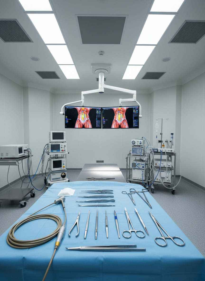
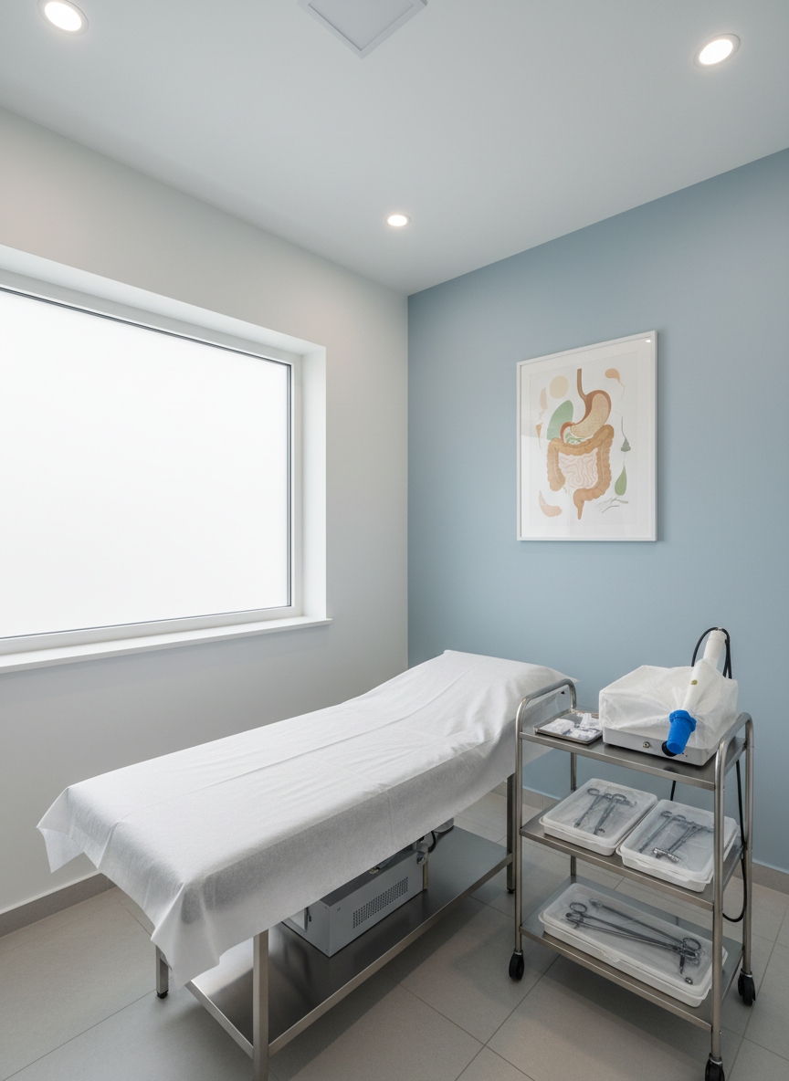
Dr Sharad Gurav is renowned laparoscopic Surgeon and Proctologist at Jupitor Hospital, Dombivli and also at Dr Gurav Surgical Clinic Dombivli. He has experience of more than 15 years in the field.He specialises in laparoscopic hernia surgery , advanced laparoscopic surgery , robotic surgery also in Laser Anorectal surgery including managment of complex anal fistula being his special interest. He has done multiple educational fellowships in laparoscopic surgery , endoscopy , Robotic Surgery and laser anorectal surgery. He has been actively involved in thousands of laparoscopic surgeries as well as endoscopy and thousands of laser anorectal surgeries since 2011. He has been practising in Dombivli area since more than 15 years and known for his compassionate and patient centric approach. He has been active in social circles via various medical camps and projects of rotary and IMA Dombivli since many years.

Piles (Hemorrhoids)

Piles are swollen and inflamed veins in the rectum and anus, commonly caused by chronic constipation, straining during bowel movements, prolonged sitting, or pregnancy. Patients may experience bleeding during defecation, pain, swelling, itching, or a prolapsing mass. Early stages can often be managed with dietary changes, increased fiber intake, medications, and lifestyle modification. Advanced or persistent cases may require procedures such as rubber band ligation, laser treatment, or surgery. Timely treatment helps prevent complications like severe pain or anemia. With modern minimally invasive techniques, treatment is safe, effective, and ensures faster recovery with minimal discomfort.

Laparoscopic hernia surgery is a modern, minimally invasive way to repair a hernia. In this procedure, the surgeon makes small cuts in the abdomen and uses a camera and special instruments to fix the weakness. A mesh is usually placed to strengthen the area and prevent recurrence. Compared to open surgery, it causes less pain, smaller scars, and faster recovery. Most patients can go home within a day and return to normal activities early. It is safe and effective when done by an experienced surgeon, but proper evaluation is important before surgery to choose the best method.

Toilet bowl filled with soil and a cactus with a pink flower

Fissure in Ano

Fissure in ano is a small tear in the lining of the anal canal, commonly caused by passing hard stools or chronic constipation. It typically presents with severe pain during and after defecation, along with occasional bleeding. Patients may also experience a burning sensation and fear of passing stools. Initial treatment includes dietary modification, stool softeners, sitz baths, and topical medications. Chronic or non-healing fissures may require procedures like lateral internal sphincterotomy or laser treatment. Early management is important to relieve pain and prevent chronicity. With appropriate treatment, most patients experience excellent relief and quick recovery.

Diagram illustrating intersphincteric, transsphincteric, suprasphincteric, and extrasphincteric anal fistulas

Fistula in Ano

Fistula in ano is an abnormal tract between the anal canal and the skin, usually developing after a poorly healed anal abscess. Patients commonly present with persistent discharge, pain, swelling, and irritation around the anus. Diagnosis is primarily clinical, though MRI may be required in complex cases. Surgery is the definitive treatment, aimed at removing the tract while preserving continence. Options include fistulotomy, laser treatment, seton placement, and advanced procedures for complex fistulas. Early treatment is important to prevent recurrence and complications. With modern minimally invasive techniques, recovery is faster and patient comfort is significantly improved.

Diagram of liver, gallbladder with gallstones, bile ducts, pancreas, and duodenum

Laparoscopic Cholecystectomy

Laparoscopic cholecystectomy is a minimally invasive surgery performed to remove the gallbladder, most commonly due to gallstones causing pain, infection, or complications. Patients typically present with upper abdominal pain, especially after meals, along with nausea or vomiting. The procedure is done using small keyhole incisions, a camera, and specialized instruments, resulting in less pain and faster recovery compared to open surgery. Most patients can go home early and resume normal activities quickly. Early surgery helps prevent recurrent attacks and serious complications, making it a safe and effective treatment option with excellent outcomes.

Anatomy of appendix including vermiform appendix, appendicitis artery, lymphoid follicles, and cross section showing lumen, lymphoid tissue, serosa

Laparoscopic Appendicectomy

Laparoscopic appendicectomy is a minimally invasive surgery performed to remove the appendix, most commonly in cases of acute appendicitis. Patients usually present with abdominal pain (often starting near the navel and shifting to the right lower abdomen), along with fever, nausea, or vomiting. The procedure is done using small keyhole incisions, a camera, and specialized instruments, resulting in less pain, minimal scarring, and faster recovery. Most patients are discharged within 24–48 hours and can return to normal activities soon. Early surgery is crucial to prevent complications like perforation or abscess, ensuring safe and effective treatment with excellent outcomes.

Laser probe treating an anal fistula with visible smoke and surgical gloves.

Laser Treatment for Pilonidal Sinus

Laser treatment for pilonidal sinus is a minimally invasive and advanced procedure used to treat infected sinus tracts near the tailbone. Patients commonly present with pain, swelling, discharge, or recurrent infections in the sacrococcygeal region. The procedure involves using a laser probe to ablate and close the sinus tract from within, without the need for large incisions. This results in minimal pain, negligible bleeding, and faster healing compared to traditional surgery. Most patients can go home the same day and resume normal activities early. Laser treatment offers excellent outcomes with low recurrence rates and improved patient comfort.

Nurse wrapping an elderly man's foot showing diabetic foot care

Diabetic Foot Management

Diabetic foot is a serious complication of diabetes resulting from neuropathy, poor blood circulation, and infection, often leading to non-healing ulcers. Early evaluation and a multidisciplinary approach are essential to prevent limb loss. Management begins with strict blood sugar control, wound care, debridement of infected or dead tissue, and appropriate antibiotics. Offloading techniques help reduce pressure on the affected area and promote healing. Vascular assessment is crucial, and revascularization procedures may be required in cases with poor blood flow. Advanced therapies such as negative pressure wound therapy and growth factor dressings can aid recovery. In complex or non-healing wounds, plastic surgery interventions like skin grafting or flap coverage may be necessary to achieve durable wound closure. Timely and comprehensive treatment significantly reduces the risk of amputation, improves healing outcomes, and helps patients return to normal function with better quality of life.

Leg with large bulging varicose veins visible on skin surface

Laser Treatment for Varicose Veins

Laser treatment for varicose veins is a minimally invasive and effective procedure used to treat enlarged, tortuous veins, commonly affecting the legs. Patients often present with pain, heaviness, swelling, visible veins, or skin changes. The procedure, known as Endovenous Laser Ablation (EVLA), involves inserting a laser fiber into the affected vein to seal it from within, redirecting blood flow to healthy veins. It is performed under local anesthesia with no large incisions, resulting in minimal pain and excellent cosmetic results. Most patients can walk immediately and resume normal activities within a day. Early treatment helps prevent complications such as ulcers or thrombosis. Laser therapy offers safe, precise, and long-lasting results with quick recovery and minimal discomfort. Check out our paper published in indexed journal on Dr Gurav’s Modification of EVLT – https://ijmedph.org/Uploads/Volume16Issue1/545.%204052.%20IJMEDPH_Sharad%20G_3185-3188.pdf

Surgeons operating in modern operation theatre

Care regarding all General Surgical Disease and conditions

A meticulously organized, state-of-the-art laparoscopic surgery workstation in a spotless, modern operating theatre, shown without any people. Stainless-steel laparoscopic instruments with fine, polished tips rest on a sterile blue drape over a smooth operating table. In the background, dual high-definition monitors display a clear, colorful 3D rendering of the lower abdomen and anorectal anatomy. Cool, diffused ceiling-mounted surgical lights cast even, shadow-free illumination, creating subtle reflections on metal surfaces. The mood is professional, calm, and reassuring. Photographed at eye level with sharp focus throughout, the composition follows the rule of thirds, emphasizing the precision tools and glowing monitors. The photographic realism and clean, minimalist aesthetic highlight advanced technology and clinical expertise appropriate for a laparoscopic and laser anorectal surgery specialist.

Treatment Costs

Flexible fee ranges and packages help you plan treatment confidently, enquire for more

About

Safe, Comfortable Surgical Experience

From your first consultation to recovery, we follow strict pre‑operative checks, modern anesthesia monitoring, and personalized post‑operative instructions, helping you heal safely with timely follow‑ups.

A pristine, modern consultation room dedicated to laparoscopic and laser anorectal care, carefully staged without any people. At the center is a cushioned examination couch with crisp white paper, adjacent to a compact trolley neatly arranged with sealed, sterile laparoscopic instrument trays and a covered laser handpiece dock. The walls are painted in soft shades of white and pale blue, with a framed, stylized illustration of the digestive tract adding subtle color. A large window with frosted glass allows gentle daylight to wash across the room, balanced by understated ceiling lights, creating a bright yet soothing ambience. Captured from a slightly elevated angle, the entire room is in sharp focus, emphasizing cleanliness, organization, and professionalism in a photographic, contemporary style.